

What is knee osteoarthritis?
The ends of the bones in your knee are covered with articular cartilage, a smooth, low-friction surface that lets the joint glide. In osteoarthritis that cartilage thins and wears, the space between the bones narrows, the bone underneath thickens and small bony spurs form at the edges of the joint. The lining of the joint can become inflamed and produce extra fluid.
It develops slowly, usually over years. It is more common with age, and it is more likely if you have had a previous knee injury or surgery, if there is a family history, if the knee is bowed or knock-kneed, or if you carry extra weight.
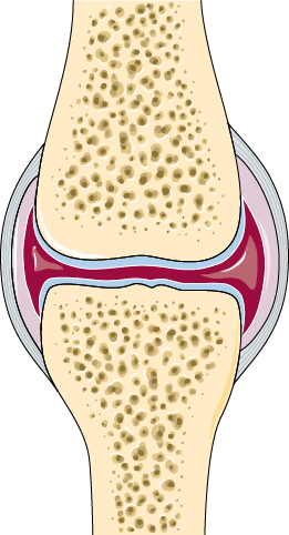
Healthy joint
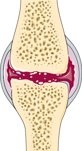
Osteoarthritis
What are the symptoms?
- Pain, usually worse with activity and eased by rest, at least at first
- Stiffness after sitting still, and first thing in the morning
- Swelling, which may come and go
- Grinding, catching or a feeling that the knee is not moving smoothly
- Difficulty with stairs, kneeling, squatting or getting out of a low chair
- In more advanced arthritis, pain at night and a knee that is becoming visibly bowed
Not all knee pain is arthritis. Meniscal tears, ligament problems, referred pain from the hip or back, inflammatory arthritis and infection all present differently and are treated differently, which is part of what an assessment sorts out.
How is it assessed?
Assessment starts with your history and an examination. Weight-bearing X-rays are the standard imaging for arthritis, because the loss of joint space is best seen with your weight through the knee. An MRI is not usually needed to diagnose osteoarthritis, though it is sometimes useful when the picture is unclear or another problem is suspected.
Importantly, how bad the X-ray looks and how bad you feel do not always match.
Treatment is guided by your symptoms and how the knee affects your life, not by the X-ray on its own.
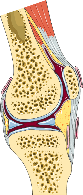
What the X-ray is actually measuring
Cartilage itself does not appear on an X-ray. What shows is the gap between the bones, and cartilage is what holds that gap open. So when a report describes joint space narrowing, it is describing the cartilage indirectly, by measuring the space it is no longer filling.
That is also why a weight-bearing film matters. Standing loads the joint and closes the space that lying down leaves open, so it shows the joint as you actually use it.
What can be done without surgery?
Most people with knee arthritis are managed without an operation, often for a long time. The measures with the best evidence behind them are unglamorous but genuinely effective for many people:
- Exercise and strengthening. Particularly the quadriceps and hip muscles. Guided physiotherapy is more effective than being told to exercise.
- Weight management, where relevant. Reducing load through the knee reduces symptoms and is one of the few things that changes the trajectory of the disease.
- Activity modification. Swapping some high-impact activity for cycling, swimming or walking, without giving up activity altogether.
- Simple pain relief, discussed with your GP or pharmacist.
- Walking aids, footwear changes and, for some, a brace.
- Injections. Corticosteroid injections can settle a flare for a period. Their effect is temporary and they are not a long-term solution.
Arthroscopy (keyhole surgery) is not a treatment for osteoarthritis of the knee. It has a role in specific mechanical problems, but the evidence does not support it as a treatment for arthritic knee pain.
When does knee replacement become a consideration?
Replacement is considered when the arthritis is advanced, the symptoms are significantly affecting your life, and non-surgical treatment is no longer holding them. There is no age at which it becomes automatic and no X-ray appearance that mandates it. It is a decision made together, based on how much the knee is costing you and what you want to be able to do.
Coming to an appointment does not commit you to surgery. A consultation may well conclude with a plan to keep going as you are, a referral for physiotherapy, or a decision to review the knee again in a year. See knee replacement for what the operation involves if that time comes.